Saturday, March 30, 2013

Life History of the Malay Tailed Judy

Life History of the Malay Tailed Judy (Abisara savitri savitri)


Butterfly Biodata:
Genus: Abisara C. & R. Felder, 1860
Species: savitri C. & R. Felder, 1860
Subspecies: savitri C. & R. Felder, 1860
Wingspan of Adult Butterfly: 40-50mm
Caterpillar Local Host Plants: Embelia ribes (Myrsinaceae), Embelia canescens (Myrsinaceae).


Physical Description of Adult Butterfly:
Above, the wings are rusty brown with two diffuse white transverse stripes on the forewing, the inner one being more sullied and stretching from mid-costa to the dorsum before the tornus. The hindwing has a similar diffuse white postdiscal band. There are two black marginal spots in spaces 4 and 5 on the hindwing separated by an orange bar. A long, white-tipped tail is present at the end of vein 4 on the hindwing. Underneath, the wings are similarly marked as per above but are more distinctly coloured with bright shades of white in the stripes set against a ground colour of yellowish brown. The basal halves of the wings are in a paler shade of yellowish brown.


Field Observations of Butterfly Behaviour:
The adults are moderately rare and are typically sighted in the forested areas of the nature reserves, and in a hill park where its host plant is growing in relative abundance. The timid and skittish adults are often seen perching on leaves with half open wings, turning and hopping from one perch to the next.


Early Stages:
The local host plants know to date are both Emelia species growing in the nature reserves. One of them, Emelia ribes, is more widespread and can also be found in various hill parks and wastelands. The immature stages of the Malay Tailed Judy feed on the relatively young leaves of the host plant. In the first two instars, the caterpillar feeds by grazing on the leaf surface while in the later instars, it feeds by chomping away at the leaf edges. Between feeds, the caterpillars of all instars rest on the leaf underside.

Local host plant #1: Embelia ribes.

Local host plant #2: Embelia canescens.

Each egg is laid singly on the underside of a leaf on the host plant, typically close to the leaf edge. Each egg is dark purplish blue, somewhat conical in shape with a diameter of about 0.75-0.8mm. The egg surface is generally smooth and there is a mid-level belt of fine hairs encircling the egg.

Two views of an egg of the Malay Tailed Judy.

A sequence of three pictures, ordered from left to right, showing the development of an egg over the course of 3 days.

A newly hatched caterpillar, next to its half eaten egg shell.

It takes about 3.5-4 days for the egg to hatch. The young caterpillar consumes part of the egg shell to emerge. With a length of about 2.0mm, it has a greyish white body with a dorsal green band. The head is greyish white. There are moderately long black dorso-lateral setae and exceptionally long whitish sub-spiracular setae. The body color changes to pale yellow with a green undertone as it feeds and grows.

Two views of a 1st instar caterpillar, late in this stage, length: 3mm

The first instar lasts for about 3 days and the body length reaches about 3.6mm. Prior to the moult to 2nd instar, the body shortens and takes on a pumped up appearance. This shortening routine also occurs prior to each of the subsequent moults.

Two views of a 1st instar caterpillar, late in this stage, length: 3mm.

The body color of the 2nd instar caterpillar is pale yellow with a greenish undertone. There are numerous very short white setae covering the body surface. The exceptionally long white sub-spiracular setae are still present and a few black setae can be found hidden among the white ones. This instar lasts for albout 3-4 days and the caterpillar grows to a length of about 6mm before the moult to the 3rd instar.

Two views of a 2nd instar caterpillar, length: 4.8mm.

Two views of a 2nd instar caterpillar, late in this stage. length: 5.3mm.

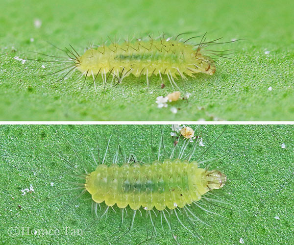
The 3rd instar caterpillar is still yellowish green but with green dominating. Otherwise, it bears a strong resemblance to the 2nd instar caterpillar. Closer scrutiny shows that no black setae are present among the sub-spiracular tuffs of white setae. After 3-4 days in this stage with the body length reaching a maximum of about 9.5-10mm, the caterpillar moults to the 4th (and penultimate) instar.

A newly moulted 3rd instar caterpillar, eating its exuviaa, length: 5.8mm.

Two views of a 3rd instar caterpillar, length: 7mm.

A 3rd instar caterpillar, dormant prior to its moult, length: 9mm

A Malay Tailed Judy caterpillar moults from the 3rd to the 4th instar.

The 4th instar caterpillar resembles the 3rd instar caterpillar but with a much denser set of sub-spiracular setae. The body is mainly yellowish green in color. The numerous short setae carpeting the body surface are now much shorter and yelow to green in colour. The head, which is colored greyish white in the first three instars, is now pale yellowish green. The 4th instar lasts for about 3.5-5 days and the body grows up to a length of about 16-16.5mm.

A newly moulted 4th instar caterpillar, next to its exuvia, length: 9mm.

Two views of a 4th instar caterpillar, length: 14mm.

A close-up view of the body surface of a 4th instar caterpillar.

A 4th instar caterpillar, dormant prior to its moult, length: 15.5mm.

The 5th instar caterpillar resembles the 4th instar caterpillar closely. The numerous short setae carpeting the body surface are now predominantly green in colour and less conspicuous. Overall the body colour is in a stronger shade of yellowish green to lime green, with the head capsule also taking on a greenish tinge.

A newly moulted 5th instar caterpillar, eating its exuvia, length: 15.2mm.

Two views of a 5th instar caterpillar, length: 26mm.

Two views of a 5th instar caterpillar, length: 29mm.

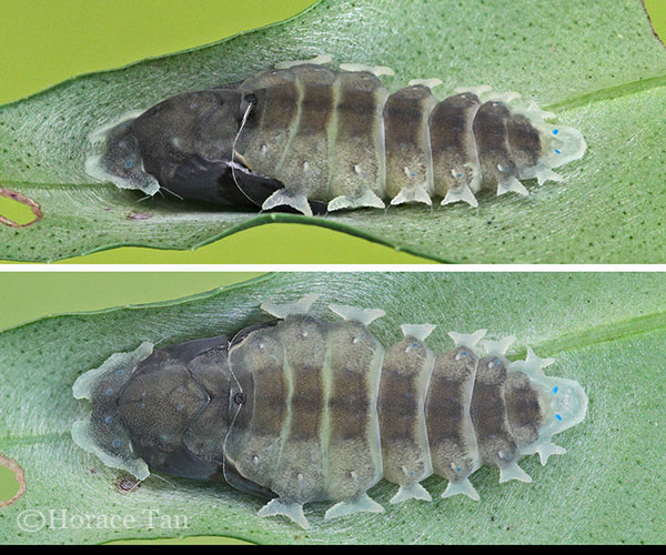
The 5th instar lasts for about 5-7 days and the body grows up to a length of about 27-29mm. On the last day of this final instar, the caterpillar ceases food intake and its body shrinks in length. It then finds a spot on the leaf underside where it spins a silk pad and a silk girdle to secure itself in a head-up manner. As the pre-pupa lays dormant, the development within causes several bluish spots and one black dorsal spot to be visible towards the end of the pre-pupal period.

Two views of a pre-pupatory larva of the Malay Tailed Judy

Pupation takes place after 1-1.5 days of the pre-pupal period. The greenish pupa has a diamond-shaped outline, being broader at mid-body, less so at the anterior end, and rather pointed at the posterior end. The body has one prominent black dorsal spot on the 1st abdominal segment and a number of bluish spots, varying from two to eight, on the remaining segments. Laterally, there are lateral wedge-shaped appendages on 3rd-9th abdominal segments. The prothorax also features lateral fin-like appendages. Each pupa is about 21-23mm in length.

A Malay Tailed Judy caterpillar moults to its pupal stage.

Two views of a pupa of the Malay Tailed Judy.

Seven days later, the pupa becomes darkened in color signaling the imminent emergence of the adult. The next day the adult butterfly emerges from the mature pupa.


Two views of the mature pupa of a male Malay Tailed Judy.

A newly eclosed Malay Tailed Judy drying its wings near its pupal case.

References:
  • [C&P4] The Butterflies of The Malay Peninsula, A.S. Corbet and H.M. Pendlebury, 4th Edition, Malayan Nature Society.
  • Butterflies of Thailand, Pisuth Ek-Amnuay, 2nd Edition, 2012
  • A Field Guide to the Butterflies of Singapore, Khew S.K., Ink On Paper Communications, 2010.
Text by Horace Tan, Photos by Benjamin Yam, Nelson Ong, Sunny Chir and Horace Tan
You have read this article with the title March 2013. You can bookmark this page URL http://butterflymuse.blogspot.com/2013/03/life-history-of-malay-tailed-judy.html. Thanks!
Tuesday, March 26, 2013

Random Gallery - Cycad Blue

Random Butterfly Gallery
The Cycad Blue (Chilades pandava pandava)


This little butterfly is common, especially in the vicinity of its caterpillar host plant, Cycas revoluta and other Cycad species. It can be found in urban parks and gardens, and in areas where various Cycads are planted - even in the nature reserves. At the main entrance of the Singapore Botanic Gardens, where two large trees of Cycas rumphii are present, the Cycad Blues are regularly seen fluttering around the plants, chasing each other in rapid flight.

This shot of a pristine Cycad Blue was taken last weekend by ButterflyCircle member Tea Yi Kai. The butterfly is perched on the edge of a Nepenthes tendril in Yi Kai's suburban garden. As long as pesticides are not used in urban parks and private gardens, butterflies are able to survive for the enjoyment of nature enthusiasts. We have often considered the wanton spraying of pesticides as 'weapons of mass destruction' because not only do these pesticides kill mosquitoes (albeit inefficiently) they destroy a large part of our urban biodiversity in the process!


You have read this article with the title March 2013. You can bookmark this page URL http://butterflymuse.blogspot.com/2013/03/random-gallery-cycad-blue.html. Thanks!
Saturday, March 23, 2013

Butterfly Bubbles

Butterfly Bubbles
Formation of bubble droplets when a Butterfly "pees"


When I was thinking of a title for this blog article, I had originally thought of a more catchy "Bubble Butts" or something to that effect.  However, a quick Google uncovered a series of rather uncomplimentary definitions that this particular pair of words alluded to, so I had to decide on a more decent title, lest this blog be subject to a more stringent censorship scrutiny!


Over the years of photographing butterflies, particularly those that were puddling, ButterflyCircle members have come across the puddlers forcefully excreting liquid from the business-ends of their abdomens. When puddling, it is quite obvious that butterflies take in copious amounts of mineral-enriched fluids that they require for various biological functions.


Jet-propelled FiveBar Swordtail!  The butterfly ejects a stream of fluids whilst puddling

The biological processing of these fluids within the bodies of the butterflies to extract the requisite minerals must be extremely efficient. Once the minerals are absorbed, the remaining fluids are ejected from the butterfly's body, so that there is no superfluous build-up of the excess fluids that the butterfly does not need.


The power of high speed photography -  A stream of droplets ejected from a puddling FiveBar Swordtail

However, it has been observed that in many cases, the excretion of the waste fluids is much slower, as the butterfly passes the fluids slowly, creating a droplet of fluid that remains attached to the tip of the abdomen of the butterfly. Given the viscosity of the liquid that the butterfly excretes, and the low speed at which the liquid forms, a droplet is formed.


Before we delve into the physics of the formation of drops of liquid, let us first recall the reasons why butterflies puddle.  In an earlier blog article, puddling was discussed. Adults of many butterflies, principally males, frequent mud puddles, stream banks that have been contaminated with animal excretions, carrions and bird droppings where their imbibe sodium, chlorides and nitrates that are essential to their biological functions. A study showed that virgin females have reduced longevity. This is attributed to virgins not obtaining important nutrients which the males transfer to females during mating.


When settled down to puddle, some butterflies can remain still for long periods of time, often to their own detriment, as they become vulnerable to nearby predators. At times, their preoccupation with puddling can prove fatal. We have observed, for example along forest road in Malaysia, where puddling butterflies are so engrossed in puddling, that they even forget to fly out of the way of an oncoming vehicle's tyres!


A Burmese Batwing forms a little bubble of fluid (taken in Thailand)

Butterflies from the families Papilionidae, Pieridae, Lycaenidae, Hesperiidae, and to a lesser extent, Nymphalidae, engage in puddling activities. In many instances, the process of excreting the excess fluids from the abdomen has been photographed. Besides the jets of fluid excreted, we have often observed the formation of small bubbles of fluid oozing out from the tip of the abdomens of butterflies.


In physics, a drop is a small column of liquid, bounded completely or almost completely by free surfaces. A drop may form when liquid accumulates at the lower end of a tube or other surface boundary, producing a hanging drop called a pendant drop.  When a butterfly excretes the liquid that flows slowly from its anal tube, the liquid forms a drop due to surface tension. A simple way to form a drop is to allow liquid to flow slowly from the lower end of a vertical tube of small diameter.


Source : Wikipedia

The surface tension of the liquid causes the liquid to hang from the tube, forming a pendant. When the drop exceeds a certain size it is no longer stable and detaches itself. The falling liquid is also a drop held together by surface tension. As the drop detaches itself from where it previously hung, surface tension will pinch it and the drop falls as a sphere.


Top : A drop of water falls off after exceeding a certain size when gravity exceeds surface tension forces. (© Wikipedia) Bottom : An Eurema simulatrix excretes a drop of fluid from its abdomen

In butterflies, it is interesting to observe the variations of the size of the drops of excreted fluids before they detach from the anal tube. There doesn't appear to be any direct relation of the size of the droplet to the size of the butterfly. In many cases, the smaller butterflies hold much larger droplets in relation to the size of the butterfly.


The drop of fluid can vary in clarity from crystal clear and totally transparent, to a muddy and cloudy constituency. This is probably dependent on the fluids that the butterfly is feeding on, or the process of digestion that the fluids go through in the alimentary canal of the butterfly.


Two examples of slightly cloudy/murky drops of excretion, compared to the usually crystal clear drop

When feeding on bird droppings, the Hesperiidae exhibit an interesting phenomenon of "recycling" their excretion.  With its proboscis extended whilst feeding on a bird dropping, a skipper has often been observed to arch its abdomen and deposit a drop of its own excreted fluid onto the bird dropping and then feeding off the mixture. It repeats the process as it adds its own excretions to the bird dropping to wet it, and then feeds for extended periods of time in this manner, if not disturbed.


Skippers recycling their pee onto the bird dropping that they are feeding on

Amongst the puddlers, butterfly species from the families Papilionidae, Pieridae, Lycaenidae and Hesperiidae tend to display the "bubble drop" phenomenon as they puddle. Of those Nymphalidae that do puddle, we have rarely come across any of the species retaining a bubble at its abdomen. Could it be perhaps that they imbibe smaller amounts of fluid as they puddle? Or maybe they have a slower metabolic process that retains in their bodies, most of the fluids that they imbibe?


In the meantime, keep observing and recording any interesting notes that could yield a better understanding of this simple phenomenon and how each different species of butterfly displays different behaviour. Click on the individual photos for a larger view of these bubbles on the butterflies.

Text by Khew SK : Photos by Chng CK, Antonio Giudici, Khew SK, Koh CH, Loke PF, Lemon Tea, Anthony Wong & Mark Wong


You have read this article with the title March 2013. You can bookmark this page URL http://butterflymuse.blogspot.com/2013/03/butterfly-bubbles.html. Thanks!